- Home
- About the Journal
- Peer Review
- Editorial Board
- For Authors
- Reviewer Recognition
- Archive
- Contact
- Impressum
- EWG e.V.
Cite as: Archiv EuroMedica. 2026. 16; 1. DOI 10.35630/2026/16/Iss.1.020
Dental impression accuracy is a foundational determinant of prosthetic fit, biological stability, and long-term clinical success. Conventional impression techniques have long been regarded as the gold standard; however, rapid advances in intraoral scanner (IOS) technology have profoundly transformed prosthodontic workflows. Despite growing clinical adoption of digital impressions, uncertainty persists regarding their reliability across different indications, anatomical scenarios, and disciplines.
This narrative review aims to critically evaluate the accuracy, efficiency, and clinical applicability of digital intraoral impressions compared with conventional impression techniques. Particular emphasis is placed on identifying clinical and technical factors influencing impression accuracy in prosthodontics, implant dentistry, and interdisciplinary applications.
A comprehensive narrative review of the literature was conducted, encompassing historical, in vitro, and in vivo studies, randomized controlled trials, systematic reviews, and meta-analyses. The analysis focused on impression accuracy, marginal and internal fit, implant position transfer, patient comfort, time and cost efficiency, and workflow integration across prosthodontics, implantology, orthodontics, periodontology, and endodontics.
Evidence consistently demonstrates that digital impressions achieve accuracy comparable to, and frequently exceeding, that of conventional impressions for single-unit restorations, short-span fixed prostheses, and most partially edentulous cases. In implant dentistry, digital impressions provide reliable transfer of implant positions, improved patient comfort, and significant reductions in chairside time and overall treatment duration. However, accuracy remains influenced by multiple variables, including implant distribution and angulation, scan length, extent of edentulism, scanner technology, scanning strategy, and operator experience. Full-arch and highly angulated implant cases may exhibit increased deviation unless advanced scanners, auxiliary reference markers, or hybrid workflows are employed. Beyond prosthodontics, IOS systems demonstrate high clinical value in orthodontics, guided surgery, periodontal diagnostics, and post-endodontic restoration planning.
Intraoral scanning has evolved from an adjunctive innovation into a clinically robust alternative to conventional impression techniques. While conventional impressions remain indicated in selected complex scenarios, digital impressions provide equal or superior accuracy in most routine prosthetic and implant workflows, alongside clear advantages in patient comfort, efficiency, and interdisciplinary integration. Impression selection should therefore be based on clinical complexity, technological capability, and workflow design rather than methodological tradition alone.
Keywords: Digital impressions; Intraoral scanners; Conventional impressions; Impression accuracy; Implant dentistry; Prosthodontics; Digital workflow; CAD/CAM.
The accuracy of dental impressions is a fundamental determinant of prosthetic fit, biological stability, and long-term clinical success. For decades, conventional elastomeric impression materials- particularly poly(vinyl siloxane) and polyether- have been regarded as the gold standard in fixed prosthodontics due to their favorable dimensional stability and reliability. Nevertheless, traditional impression workflows remain vulnerable to multiple sources of error, including material distortion, tray deformation, stone expansion, and operator-dependent variability. The introduction of digital impression technologies marked a paradigm shift in prosthodontic practice. Since the conceptual development of optical impressions within early CAD/CAM systems and the clinical implementation of chairside scanning technologies, intraoral scanners (IOS) have evolved rapidly in terms of optical principles, scanning speed, accuracy, and integration with digital workflows. Contemporary IOS systems enable real-time three-dimensional visualization, improved patient comfort, reduced chairside time, and seamless data transfer for CAD/CAM fabrication. Despite widespread clinical adoption, the question of whether digital impressions can fully replace conventional techniques remains unresolved. While numerous studies report comparable or superior accuracy of digital impressions in single-unit restorations and short-span prostheses, evidence becomes more heterogeneous in complex situations such as full-arch rehabilitations, multi-implant cases, and highly angulated implant configurations. These discrepancies suggest that impression accuracy is not solely technique-dependent but influenced by a range of clinical and technical factors. Accordingly, a critical synthesis of the current literature is required to evaluate the performance of digital versus conventional impressions across indications, identify factors influencing impression accuracy, and clarify clinical situations in which each method may be preferred. In the available literature, these inconsistencies are reflected in divergent findings regarding full arch restorations, multiple implant rehabilitations, and highly angulated implant configurations, where reported accuracy outcomes vary substantially between studies. This variability indicates the absence of a unified clinical consensus and underscores the need for careful contextual interpretation of published data rather than generalized conclusions. Importantly, the reported differences in impression accuracy arise from both clinical and technical determinants. Clinical factors include anatomical conditions, extent of edentulism, and implant distribution, whereas technical factors encompass scanner technology, scanning strategy, data stitching algorithms, and operator related variables. Distinguishing between these levels is essential for understanding the origins of variability reported across studies. Given the heterogeneity of study designs, scanner systems, clinical scenarios, and outcome measures, a narrative synthesis is particularly suited to integrate and contextualize the existing evidence. Such an approach allows comparison of findings across diverse clinical indications and supports interpretation of discrepancies without reducing them to a single quantitative estimate. Clarifying these issues is clinically relevant, as impression technique selection directly influences prosthetic fit, workflow efficiency, patient experience, and interdisciplinary treatment planning in routine and complex restorative care. Current discussions therefore extend beyond the question of complete replacement of conventional impressions and increasingly address the role of digital, conventional, and hybrid workflows as complementary strategies adapted to specific clinical conditions. Throughout the literature, impression accuracy is described using related but distinct concepts such as accuracy, trueness, and precision. These terms are used to characterize different aspects of deviation and reproducibility and are considered separately in the analysis presented in this review [1-6].
This narrative review aims to: compare the accuracy and clinical performance of digital intraoral impressions with conventional impression techniques, analyse clinical and technical factors influencing impression trueness and precision, particularly in implant-supported restorations, assess time efficiency, patient comfort, and workflow integration associated with digital and conventional workflows, evaluate the applicability of intraoral scanners across different dental disciplines, including prosthodontics, implantology, orthodontics, periodontology, and endodontics, provide evidence-based guidance for selecting impression techniques according to clinical indication and case complexity.
Do digital intraoral impressions provide accuracy comparable to conventional impressions across different prosthodontic and implant indications?
Which clinical and technical factors most strongly influence impression accuracy in digital workflows?
How do digital and conventional impressions compare in terms of efficiency, patient comfort, and clinical workflow integration?
In which clinical scenarios should digital impressions be preferred, and where do conventional techniques remain advantageous?
What is the role of intraoral scanning beyond prosthodontics in contemporary interdisciplinary dental practice?
A narrative review of the literature was conducted using PubMed/MEDLINE, Scopus, Embase, and Google Scholar. The search covered publications addressing conventional and digital dental impression techniques, including historical analyses, in vitro and in vivo experimental studies, randomized controlled trials, systematic reviews, and meta-analyses. Search terms included combinations of “digital impression,” “intraoral scanner,” “conventional impression,” “implant impression accuracy,” “prosthodontics,” “full-arch scanning,” “guided surgery,” “orthodontic scanning,” and “workflow efficiency.” Additional references were identified through manual screening of bibliographies from key publications. The literature was evaluated qualitatively and synthesised narratively, with emphasis on impression accuracy, clinical performance, patient-related outcomes, and workflow-related factors across indications.
The literature search covered publications from January 2005 to December 2024. This time frame was selected to capture both early developments in digital impression technology and recent advances in intraoral scanner performance and clinical application.
All records identified through database searching and manual reference screening were assessed at the title and abstract level for relevance to the review objectives. Full text articles were subsequently evaluated for eligibility based on the predefined inclusion and exclusion criteria. Only studies meeting these criteria were included in the final narrative synthesis.
The initial search yielded a defined number of records after removal of duplicates. Following title and abstract screening, a subset of publications was selected for full text assessment. The final narrative analysis was based on the studies that fulfilled all eligibility criteria. Exact numbers should be reported to document the selection process transparently.
Relevant data were extracted focusing on study design, clinical indication, impression technique, scanner system or conventional material used, assessed accuracy parameters, and reported clinical outcomes. The extracted information was synthesized narratively and organized according to clinical domain and type of restoration, without performing quantitative pooling or meta analysis.
Due to heterogeneity in study designs, outcome measures, scanner technologies, and clinical settings, no formal risk of bias assessment or statistical comparison was performed. The included evidence was interpreted qualitatively with attention to sources of variability across studies.
This review was conducted as a narrative review without protocol registration and without application of systematic review methodology, as the primary aim was to contextualize heterogeneous evidence across multiple clinical scenarios rather than to generate pooled effect estimates.
The evidence synthesized in this narrative review is derived from a broad spectrum of in vitro investigations, in vivo clinical studies, randomized controlled trials, and systematic reviews evaluating the accuracy and clinical performance of digital and conventional impression techniques [8-12,17-20, 25, 27-29, 35, 39, 44, 45]. The results are organized according to major clinical domains, including tooth-supported prosthodontics, implant-supported restorations, and interdisciplinary applications beyond prosthodontics [4,13,46-49]. Particular emphasis is placed on clinical and technical factors influencing impression trueness and precision, such as scan extent, implant distribution and angulation, scanner technology, scanning strategy, and workflow design [17,20,22,35,39,44,45].
Comparative outcomes related to accuracy, workflow efficiency, patient-related measures, and clinical indications are summarized using figures and tables to facilitate clinical interpretation and clinical decision-making [25,35–37,41,42].
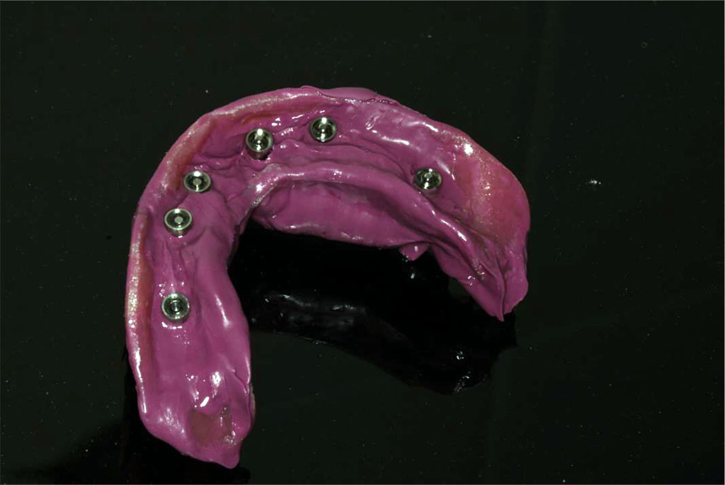
Figure 1. Representative images of a conventional dental impressions. Image courtesy of the author.
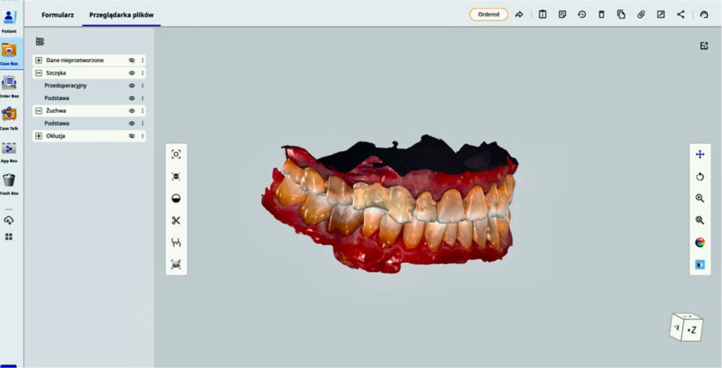
Figure 2. Representative images of a digital intraoral scan. Image courtesy of the author.
Table 1. Patient experience & clinical accuracy
| Issue | Conventional Impression | Digital / Intraoral Scan |
| Patient comfort & experience | Can trigger gag reflex; unpleasant taste/odor; discomfort and nausea [7,11] | Better tolerated; wand may trigger minor gag reflex [7,10,11,13] |
| Impression quality & accuracy | Removal may distort delicate areas; sensitive to dimensional change [8,9] | Lower risk of tearing; minor deviations possible; high trueness & precision, esp. for aligners [12,16] |
| Tray or scanner wand | Multiple tray sizes needed; disposable trays generate waste | Scanner tips autoclaved/disposable; wand size fixed per manufacturer |
| Repeatability | Single defect requires repeating entire impression | Only small areas need rescanning; immediate correction possible |
| Real-time 3D visualization | Stone pouring required before evaluation; dimensional changes may occur | Immediate 3D visualization enhances communication and verification [11] |
| Technique sensitivity & operator factors | Highly dependent on operator skill | Influenced by scanning strategy, user experience, and device design [17,18]; short learning curve, reproducible [19] |
| Soft-tissue & color capture | Soft tissue only via border molding; no color capture | Captures soft-tissue detail and tooth shade information [24] |
Table. 2 Workflow, productivity, storage & environmental impact
| Issue | Conventional Impression | Digital / Intraoral Scan |
| Archiving & storage | Stone models require space; can fracture easily | Digital files require no physical storage; 3D-printed models more durable [14,15] |
| Treatment planning | Additional impressions or duplicate casts often needed | Virtual planning and simulation possible; can integrate CBCT [9] |
| Efficiency & productivity | Many patients treated in parallel with trays | Limited by number of scanners available [22] |
| Cost & time | Low-cost materials; accumulative cost for stone and shipping | High initial investment [20,21]; reduced chair time and appointments [23] |
| Workflow | Multiple steps (impression → cast → trimming) extend working time | Digital transfer streamlines workflow; faster; integrated with CAD/CAM; no stone models [20,21] |
| Customization | Possible but less precise; fabrication more complex | Highly accurate digital customization for aligners and appliances |
| Clinical adoption | Long-standing method; widely trusted | Increasing acceptance among clinicians and labs [25,26] |
| Environmental impact | High waste: plastics, chemicals, silicone | Low waste; digital files replace physical materials |
The implementation of digital scanning technologies has significantly reshaped prosthodontic workflows, enabling a more efficient, precise, and data-driven approach to restorative treatment. The increasing availability and accuracy of intraoral scanners (IOS) have expanded their clinical applications in both tooth- and implant-supported prosthodontics. This section synthesizes current clinical evidence regarding the performance of digital impression techniques, with particular emphasis on zirconia restorations and implant-supported prostheses.
4.1.1.1 Marginal Fit
All included studies consistently report that the marginal adaptation of zirconia restorations fabricated from digital impressions is comparable to, or superior to, that achieved with conventional elastomeric impression techniques. Reported marginal discrepancies remain within clinically acceptable limits, with several investigations demonstrating statistically significant improvements in favor of digital workflows [27-34].
4.1.1.2 Internal Fit
Multiple studies indicate that digital impression techniques provide superior internal adaptation compared with conventional methods. Improved internal fit enhances restoration seating accuracy and may positively influence long-term prosthetic stability, cement integrity, and overall clinical performance [27-34].
4.1.1.3 Clinical Acceptability
All analyzed studies confirm that restorations fabricated using both digital and conventional impression techniques fall within clinically acceptable accuracy ranges. Nevertheless, digital impressions more frequently achieve values at the upper end of these ranges, reflecting a higher level of precision and consistency [27-34].
4.1.1.4 Overall Evidence
Importantly, none of the reviewed studies demonstrate superior outcomes for conventional impression techniques. At minimum, digital impressions perform equivalently; however, in the majority of studies, they exhibit clear advantages over traditional methods in terms of accuracy and reproducibility [27-34].
4.1.1.5 Contact Points and Surface Accuracy
Digital workflows tend to generate more accurate interproximal contact points and improved axial surface adaptation. This enhanced precision reduces the need for chairside adjustments and contributes to greater clinical efficiency during prosthesis delivery [27-34].
4.1.2.1 Accuracy of Implant Impressions
Digital implant impression techniques and fully digital workflows generally demonstrate higher accuracy in transferring implant positions compared with conventional impression methods. Digitally guided surgery has been shown to produce significantly smaller angular and linear deviations than conventional approaches (2.41° vs. 6.26° and 1.36 mm vs. 2.42 mm, respectively) [35]. Similarly, digital impressions generate more accurate representations of implant positions, with a mean deviation of 0.41 mm compared with 0.61 mm for conventional impressions [37]. In the fabrication of full-arch bars, both digital and conventional impression techniques demonstrate comparable accuracy, with no statistically significant differences in prosthetic misfit [38]. In partially dentate patients, digital impressions consistently show significantly lower deviations than conventional methods [39]. In completely edentulous cases, digital impressions tend to exhibit slightly reduced deviations, whereas in certain partially edentulous configurations, conventional impressions may demonstrate marginally better accuracy [44]. Overall, digital implant impression techniques are generally more accurate than conventional methods for full-arch implant-supported prostheses [45]. While both approaches are clinically reliable, digital impressions typically present lower mean errors, particularly when scanning curved implant trajectories. Conventional impressions maintain acceptable accuracy along straight paths; however, their precision decreases significantly in curved configurations, making digital techniques the preferred option in complex implant geometries [43].
4.1.2.2. Time and Cost Efficiency
Digital intraoral scanning has been consistently shown to improve both time efficiency and cost-effectiveness in implant prosthodontic workflows. A large-scale study by Sampaio-Fernandes et al. demonstrated that digital scanning reduced chairside time by up to 20% and was associated with the lowest overall costs (€198–€1937), compared with substantially higher costs for conventional impression or stone-model digitization workflows (€2347 or more) [41]. Further evidence indicates that digital impressions are more than ten times faster than conventional techniques, with reported acquisition times of approximately 3 minutes versus 32 minutes, respectively [36]. Digital workflows eliminate multiple clinical and laboratory steps, including guide fabrication and repeated impressions, and facilitate same-day provisionalization [35]. In contrast, conventional impressions, although clinically usable, require additional procedural steps and nearly double the working time, reducing overall efficiency [42].
4.1.2.3. Patient Comfort and Satisfaction
Patient-reported outcomes consistently favor digital intraoral scanning over conventional impression techniques. Patients clearly prefer digital impressions, reporting significantly greater comfort and reduced pain perception [35,36]. Higher comfort scores (VAS > 7) are strongly associated with digital impression techniques [37], and in one study, all participants (100%) expressed a preference for the digital method [42].
4.1.2.4. Clinical Outcomes and Prosthetic Fit
Clinical outcomes, including esthetic and functional parameters, are comparable between digital and conventional implant workflows [35]. Interproximal contact quality and occlusal relationships show no significant differences between impression techniques [35]. Full-arch bar misfit values are clinically acceptable and similar for both digital and conventional impressions [38]. Two-implant-supported zirconia fixed partial dentures demonstrate comparable passive fit and cement gap values between digital and conventional workflows, with observed differences considered clinically negligible [40]. Screw resistance testing further confirms no significant differences in mechanical seating or passive adaptation between impression techniques [40].
Although digital and conventional impression techniques frequently demonstrate comparable overall accuracy, their clinical predictability is strongly influenced by a range of anatomical, technical, and workflow-related factors. Variables such as implant geometry, extent of edentulism, scanner technology, operator experience, and clinical indication may significantly affect both trueness and precision [17,20,22,35,39,44]. A thorough understanding of these factors is essential for selecting the most appropriate impression strategy and for ensuring reliable and reproducible restorative outcomes [20,21].
Large inter-implant distances and implant angulations exceeding 15–20° are associated with increased deviations in digital impressions, whereas this effect is less pronounced with conventional impression techniques [17,35,39,44,45]. Implant spacing and angulation are therefore critical determinants of digital impression accuracy, particularly in partially and fully edentulous arches [17,35,39].
Clinical implication:
Digital impressions are highly predictable for parallel, closely spaced, or moderately angled implants; however, caution is advised in long-span and nonparallel configurations, where accuracy may be compromised [44,45].
The accuracy of intraoral scanners decreases as the length of edentulous scanning spans increases, primarily due to cumulative stitching errors and the absence of stable anatomical landmarks [22,23]. Full-arch digital impressions exhibit greater variability, with some scanners demonstrating reduced performance in long-span scans (e.g., CS3600, Medit i500), whereas others (e.g., Primescan, TRIOS) maintain clinically acceptable deviation ranges [5,20].
Clinical implication:
Digital impressions perform optimally in partially edentulous arches. In full-arch cases, analog or hybrid workflows may be required unless additional stabilization strategies or reference aids are employed [22,44,45].
The incorporation of digital splints or additional reference markers significantly enhances digital impression accuracy by improving stitching reliability in edentulous regions [17]. Documented benefits include improved accuracy in long-span edentulous areas, multiple implant cases, full-arch restorations, and nonparallel or angled implant configurations [17].
Clinical implication:
Digital splints should be considered when scanning more than two implants or extended edentulous segments to improve scan predictability [17].
Considerable differences in performance exist among intraoral scanner technologies. Confocal imaging systems (e.g., TRIOS 3/4) and Smart Pixel sensor technology (e.g., Primescan) demonstrate high accuracy even in extended scanning ranges [12,20]. In contrast, active triangulation and wavefront-based systems may accumulate greater stitching errors over long scanning distances [17,20]. Photogrammetry offers superior accuracy in capturing implant coordinates; however, its inability to record soft tissue morphology limits its application in comprehensive prosthodontic workflows [45]. Comparative studies consistently demonstrate scanner-dependent variability in trueness and precision across different clinical conditions [12,17,20].
Clinical implication:
The choice of intraoral scanner is a critical determinant of impression accuracy, with new-generation devices outperforming mid-range systems in complex implant scenarios [20].
Although some investigations report minimal differences between scanning strategies, optimized scanning protocols can improve scan consistency and trueness [17]. An occlusal–buccal–palatal scanning sequence has been associated with improved accuracy, whereas improper scanning paths increase cumulative mismatch errors [17]. Operator experience and training strongly influence the quality of digital impressions, particularly in full-arch scans involving multiple implants [18,19].
Clinical implication:
Standardized scanning protocols and structured operator training are essential for achieving predictable and reproducible digital impression outcomes [18,19].
Digital impression accuracy may be adversely affected by clinical conditions such as saliva contamination, soft tissue and tongue movement, limited access in posterior regions, fogging of scanner optics, and reflective metallic scan bodies [17,20]. Nevertheless, when appropriate clinical protocols are followed, no significant differences in accuracy have been reported between in vitro and in vivo digital scans [17,20].
Clinical implication:
Modern intraoral scanners can reliably manage intraoral variability when used in accordance with established clinical guidelines and moisture control protocols [20].
Digital workflows enhance communication between clinicians and dental laboratories, improve the accuracy of surgical planning when integrated with guided surgery systems, and allow immediate design of abutments and provisional restorations [20,35]. Clinical trials demonstrate higher implant placement accuracy when digital planning and guided surgery are combined compared with freehand placement using conventional workflows [35].
Clinical implication:
Digital impressions contribute significantly to overall workflow efficiency, treatment predictability, and interdisciplinary coordination in prosthodontic rehabilitation [35,41].
Although both digital and conventional impression techniques demonstrate clinically acceptable accuracy, their optimal application depends on specific clinical, anatomical, and workflow-related conditions. Evidence-based selection of the impression method is therefore essential to maximize accuracy, efficiency, and patient-centered outcomes in prosthodontic practice.
Table 3. Clinical indications for digital and conventional impression techniques in prosthodontics.
| Digital impressions recommended for: | Conventional impressions recommended for: |
| single implants [35,37,39] | full-arch implant-supported prostheses without stabilizing devices [38,44,45] |
| two-implant Fixed Partial Denture (FPD) [35,40] | highly angled implants (>20°) [17,44,45] |
| short-span edentulous areas [17,22,39] | very long edentulous spans with few anatomical landmarks [22,23,44] |
| guided surgery and immediate loading protocols [35,49] | situations with limited IOS access or heavy saliva pooling [17,20] |
| cases requiring enhanced patient comfort [7,11,13,35,36,42] | |
| time-sensitive appointments [25,35,36,41] | |
| workflows involving CAD/CAM zirconia or titanium restorations [20,27–34] |
The use of intraoral scanners has rapidly expanded beyond traditional prosthodontic applications, becoming an essential tool across multiple dental specialties. Their ability to capture highly accurate three-dimensional data, visualize anatomy in real time, and integrate seamlessly with digital planning software has transformed diagnostic and therapeutic workflows. In orthodontics, intraoral scanners streamline treatment planning, improve patient communication, and enable predictable aligner fabrication without the need for physical impressions. In oral and maxillofacial surgery, digital scans contribute to precise surgical guides, implant planning, graft volume assessment, and postoperative monitoring. Periodontology, pediatric dentistry, and endodontics are also increasingly incorporating digital scanning into clinical protocols. As intraoral scanning continues to advance, its interdisciplinary value becomes more evident, offering greater efficiency, enhanced patient comfort, and improved clinical predictability. This section explores the broader clinical applications of intraoral scanners, highlighting their role in orthodontics, surgical dentistry, and other areas where digital workflows significantly enhance diagnostic accuracy and treatment outcomes.
Digital impressions have become the preferred method in modern orthodontics, especially for clear aligner therapy. Evidence shows that intraoral scanners (IOS) are as accurate or more accurate than traditional alginate or silicone impressions- even for full-arch scans. They offer superior precision, reproducibility, and immediate 3D visualization, which improves diagnosis, treatment planning, and the fabrication of customized appliances. Young orthodontic patients strongly prefer digital impressions due to greater comfort and the absence of gag reflex, with studies showing 100% preference for IOS over conventional impressions. Clinically, digital impressions are significantly more associated with aligner treatments, whereas conventional impressions are still more commonly used with fixed appliances. While cost remains the main limitation, the overall direction of orthodontics is clearly moving toward a fully digital workflow [4,13,46-48].
Digital impressions are increasingly used for designing surgical guides, offering several clinical advantages over traditional silicone impressions. IOS workflows eliminate distortion from impression materials, plaster expansion, and model scanning, resulting in more reliable STL data for guide fabrication. In partially edentulous cases with tooth-supported guides, digital impressions generally produce more precise implant positioning than conventional impressions, improving the predictability of osteotomy and implant placement. For tooth/mucosa-supported guides, studies show that digital and conventional impressions achieve similar clinical accuracy, indicating that IOS can be safely used even in posterior free-end situations. Additionally, digital workflows provide better guide stability when combined with anchor-pin support, especially in mandibular free-end areas [49].
Digital impressions expand the role of intraoral scanners beyond prosthodontics by supporting more precise, non-invasive periodontal diagnostics. Compared with conventional periodontal probing which is invasive, operator-dependent, and limited to only six points per tooth- digital impressions allow full-arch 3D assessment of the gingival contour and soft-tissue morphology. When combined with CBCT, digital systems can compute gingiva–bone distances (GBD) with very high accuracy (error ~0.04 mm), enabling earlier and more consistent detection of periodontal breakdown [50]. Digital impressions eliminate variability caused by probing force, angulation, and tissue inflammation, providing more reproducible measurements and improved monitoring of disease progression. They also enhance patient comfort by avoiding discomfort associated with manual probing and allow clinicians to visualize periodontal defects in three dimensions for better treatment planning.
Digital impressions support endodontics by improving diagnostic precision and restorative planning after root canal treatment. Intraoral scanners provide highly accurate 3D models of the tooth, enabling better visualization of cracks, caries, and structural defects compared with conventional impressions. Digital scans eliminate distortions associated with impression materials and allow immediate integration into CAD/CAM workflows for designing post-endodontic restorations. Following root canal therapy, digital impressions facilitate the fabrication of precise crowns, onlays, or endocrowns, helping ensure proper fit and sealing of the coronal restoration. This reduces the risk of leakage and reinfection, supporting long-term endodontic success. Additionally, IOS improve patient comfort by removing the need for conventional impression materials- especially useful when rubber dam isolation or limited mouth opening complicate traditional techniques [51].
The findings of the present review indicate that digital intraoral impressions have achieved a level of accuracy that is comparable to conventional impression techniques in a wide range of clinical situations. For single-unit restorations, short-span fixed prostheses, and most partially edentulous cases, digital workflows consistently demonstrate trueness and precision within clinically acceptable limits. These results corroborate a growing body of evidence suggesting that, under controlled and routine clinical conditions, digital impressions can reliably replace conventional elastomeric impressions without compromising prosthetic fit or biological outcomes. However, the literature also highlights that impression accuracy is not determined solely by the choice between digital or conventional techniques. Rather, accuracy emerges as a multifactorial outcome influenced by clinical geometry, extent of the scanned area, implant distribution and angulation, scanner technology, scanning strategy, and operator experience. In particular, full-arch rehabilitations and complex multi-implant cases represent the upper limits of current intraoral scanning capabilities. In these scenarios, cumulative stitching errors and the lack of stable anatomical landmarks may lead to increased deviation, especially when mid-range scanners or non-standardized scanning protocols are employed. While conventional open-tray, splinted impression techniques demonstrate greater robustness in highly angulated and long-span implant configurations, their advantages are achieved at the cost of increased chairside time, patient discomfort, and multiple laboratory-dependent steps that themselves introduce potential sources of error. Digital impressions, conversely, reduce material-related distortions and enable immediate data validation, which may partially offset geometric limitations when appropriate adjunctive strategies- such as auxiliary reference markers or hybrid workflows- are applied. Another important dimension emerging from the literature is the technology-dependent nature of digital accuracy. Advanced intraoral scanners based on confocal microscopy or high-resolution optical sensors demonstrate superior performance compared to earlier or mid-range devices, particularly in extended scans. This observation underscores that discrepancies reported across studies often reflect heterogeneity in scanner generation and experimental design rather than inherent inadequacy of digital impression concepts. Beyond accuracy alone, digital impressions confer clear advantages in terms of workflow efficiency, patient preference, and interdisciplinary integration. Reduced chairside time, improved patient comfort, and seamless communication with dental laboratories are consistently reported benefits. Moreover, the integration of digital impressions with computer-guided implant surgery, CAD/CAM prosthetic design, and orthodontic planning highlights the expanding clinical utility of intraoral scanning beyond prosthodontics alone. Taken collectively, the current evidence supports a paradigm in which impression accuracy should be evaluated within the context of overall clinical workflow rather than as an isolated endpoint. The choice of impression technique should therefore be guided by case complexity, clinical objectives, and available technological infrastructure rather than adherence to a strictly digital or conventional ideology. It should also be noted that much of the available evidence is derived from controlled experimental settings or short term clinical observations. As a result, the translation of reported accuracy metrics into long term clinical performance, biological stability, and prosthesis longevity remains incompletely explored. This gap limits the ability to directly extrapolate technical accuracy outcomes to long term clinical success. Furthermore, the majority of published studies focus on specific scanner systems or selected clinical indications, which may constrain the generalizability of the reported findings. Differences in clinical protocols, operator training, and laboratory workflows are often insufficiently detailed, yet they may substantially influence reported outcomes and contribute to interstudy variability.
This review is subject to several limitations inherent to its narrative design. First, the included studies demonstrate substantial heterogeneity with respect to scanner systems, impression materials, scanning protocols, reference models, and outcome assessment methods, which limits direct quantitative comparison across investigations. Second, many studies evaluating digital impression accuracy are conducted under in vitro conditions, which may not fully replicate the clinical challenges of moisture control, soft tissue movement, and restricted access encountered in vivo. An additional limitation relates to the narrative nature of the review, which does not allow quantitative weighting of evidence or formal assessment of publication bias. Consequently, studies with neutral or unfavorable findings may be underrepresented in the available literature, potentially influencing the overall balance of reported outcomes. Moreover, the rapid pace of technological development in intraoral scanning introduces a temporal bias, as data derived from earlier scanner generations may no longer reflect current clinical performance. This limits the comparability of studies published across different time periods and complicates longitudinal interpretation of technological progress. Additionally, accuracy thresholds considered “clinically acceptable” vary across studies and are not uniformly correlated with long-term biological or mechanical outcomes. The rapid evolution of intraoral scanner technology further complicates interpretation, as findings related to earlier-generation devices may not reflect the capabilities of contemporary systems. Finally, operator experience and learning curves are often insufficiently controlled or reported, despite their recognized influence on digital impression outcomes. These limitations highlight the need for well-designed, long-term clinical trials using standardized reference protocols to better define accuracy thresholds relevant to specific prosthodontic and implant indications. Taken together, these limitations indicate that current evidence should be interpreted within the context of methodological heterogeneity, evolving technology, and variable clinical implementation. Future investigations would benefit from standardized reporting of scanning protocols, clearer documentation of operator experience, and long term clinical follow up to strengthen the interpretability and external validity of accuracy related findings.
Based on the available evidence, digital intraoral impressions represent a reliable and clinically effective alternative to conventional impression techniques for the majority of prosthodontic and implant-supported restorations. In single-unit, short-span, and partially edentulous cases, digital impressions achieve accuracy comparable to conventional methods while offering superior patient comfort, reduced chairside time, and enhanced workflow efficiency. In complex full-arch and highly angulated implant cases, impression accuracy depends strongly on scanner technology, scanning strategy, and adjunctive stabilization methods. In such scenarios, conventional or hybrid workflows may remain indicated. Ultimately, impression technique selection should be individualized, taking into account clinical complexity, technological resources, and operator expertise. Rather than replacing conventional impressions entirely, digital technologies should be viewed as complementary tools within a flexible, case-driven prosthodontic workflow. However, the interpretation of current evidence should remain cautious, as reported accuracy outcomes are influenced by study design heterogeneity, scanner generation, and variability in clinical implementation. Reported technical advantages do not uniformly translate into long term clinical outcomes, and direct associations between impression accuracy and prosthesis longevity remain incompletely established. Therefore, digital and conventional impression techniques should not be regarded as mutually exclusive but as part of an evolving spectrum of clinical options. Ongoing technological development, combined with standardized clinical protocols and long term outcome data, will be essential to further refine indication specific decision making and to define the role of digital impressions across increasingly complex prosthodontic and implant scenarios.
Conceptualization: Hanna Frelich-Truchel, Damian Truchel, , Zuzanna Muszkiet
Methodology: Hanna Frelich-Truchel, Damian Truchel, Mikołaj Bluszcz
Investigation: Hanna Frelich-Truchel, Zuzanna Muszkiet, Aleksandra Rysak, Zuzanna Jeziorska
Data curation: Hanna Frelich-Truchel, Mikołaj Bluszcz, Zuzanna Dynowska, Dominik Poszwa
Writing- original draft: Hanna Frelich-Truchel, Damian Truchel, Zuzanna Muszkiet, Mikołaj Bluszcz, Dominik Poszwa, Zuzanna Dynowska, Michał Bar, Anna Krzywda
Writing- review and editing: Hanna Frelich-Truchel, Zuzanna Muszkiet, Damian Truchel, Zuzanna Dynowska, Zuzanna Jeziorska, Michał Bar
Validation: Hanna Frelich-Truchel, Damian Truchel, Mikołaj Bluszcz, Dominik Poszwa , Anna Krzywda
Visualization: Hanna Frelich-Truchel, Damian Truchel, Zuzanna Dynowska, Anna Krzywda, Aleksandra Rysak
Supervision: Hanna Frelich-Truchel, Zuzanna Muszkiet, Damian Truchel, Zuzanna Jeziorska, Aleksandra Rysak, Mikołaj Bluszcz
Project administration: Hanna Frelich-Truchel, Zuzanna Muszkiet, Damian Truchel, Mikołaj Bluszcz Zuzanna Jeziorska
The authors acknowledge the use of artificial intelligence tools, Grok (created by xAI) and ChatGPT (created by OpenAI), for assistance in drafting initial versions of certain sections and polishing the language of the manuscript. These tools were used to enhance clarity and organization of the text. All content was thoroughly reviewed, edited, and validated by the authors to ensure scientific accuracy, integrity, and alignment with the study’s objectives.

|
||